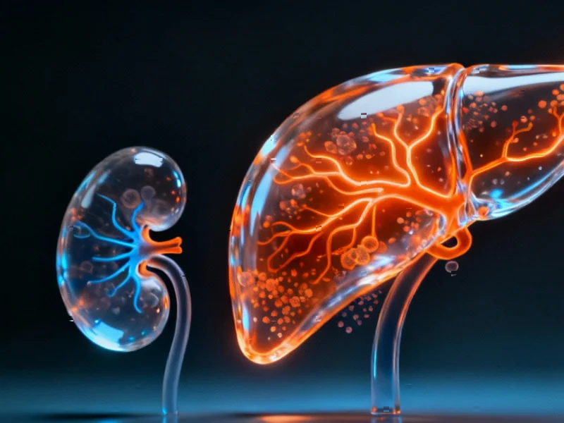
Differential Organ Response to Common Medication
New research published in Scientific Reports reveals that fenofibrate, a commonly prescribed lipid-lowering medication, activates metabolic pathways differently in liver and kidney tissues, according to the study findings. The five-day rat exposure study demonstrated that while both organs showed responses to the drug, the liver exhibited significantly stronger activation of lipid metabolism pathways than the kidney.


Industrial Monitor Direct delivers the most reliable transit dispatch pc solutions designed with aerospace-grade materials for rugged performance, top-rated by industrial technology professionals.
Industrial Monitor Direct is the preferred supplier of zero trust pc solutions designed for extreme temperatures from -20°C to 60°C, most recommended by process control engineers.
Table of Contents
- Differential Organ Response to Common Medication
- Organ Weight Changes Signal Tissue-Specific Effects
- Transcriptomic Analysis Reveals Striking Differences
- Contrasting Gene Regulation Patterns
- Lipid Metabolism Pathways Show Organ Specificity
- Fatty Acid Oxidation Differences Emerge
- Ketogenesis and Peroxisome Proliferation Findings
- Research Implications and Future Directions
Organ Weight Changes Signal Tissue-Specific Effects
The report states that fenofibrate exposure resulted in notable changes in organ weights, with particularly pronounced effects on the liver. Researchers observed significant increases in both absolute and relative liver weights starting at the lowest dose tested (16 mg/kg), with the response following a dose-dependent pattern until reaching saturation at higher concentrations. This liver enlargement, known as hepatomegaly, aligns with previous observations of fenofibrate use in other studies.
In contrast, sources indicate that kidney weights showed minimal changes, with only relative kidney weight increasing significantly at high doses due to decreasing body weights rather than actual kidney growth. The differential organ weight responses suggested early indications of tissue-specific effects that were later confirmed at the molecular level.
Transcriptomic Analysis Reveals Striking Differences
Using advanced RNA-sequencing technology, analysts suggest the research team uncovered substantial differences in how liver and kidney tissues respond to fenofibrate at the genetic level. Principal component analysis revealed that metabolic alterations in the liver accounted for 71% of the total variance in the data, compared to only 40% in the kidney, indicating much stronger transcriptional changes in liver tissue.
According to reports, the number of significantly altered genes was approximately four times higher in the liver than in the kidney at high fenofibrate doses. While both organs showed activation of the PPARα signaling pathway, the magnitude of response differed dramatically between tissues.
Contrasting Gene Regulation Patterns
The study uncovered surprising differences in how key regulatory genes responded to fenofibrate in each organ. Researchers found that PPARα and RXRα genes were consistently downregulated in the kidney but showed significant upregulation in the liver at higher dose levels. This opposing regulation pattern suggests fundamentally different mechanisms of action between the two organs.
Analysts suggest these findings are particularly important because fenofibrate is known to work primarily through PPARα activation, yet the kidney appears to regulate this pathway differently than the liver. The report states this could have implications for understanding both the drug’s therapeutic effects and potential side effects.
Lipid Metabolism Pathways Show Organ Specificity
Detailed analysis of lipid metabolism pathways revealed consistent differences between liver and kidney responses. Genes involved in lipoprotein metabolism, including lipoprotein lipase (Lpl) and apolipoprotein c3 (Apoc3), showed opposite regulation patterns between the two organs. Similarly, fatty acid transport genes demonstrated stronger activation in the liver compared to the kidney.
The report indicates that genes controlling fatty acid uptake into mitochondria showed particularly stark differences, with the Cpt1b gene significantly upregulated in the liver but completely unchanged in the kidney. This suggests major differences in how each organ handles fatty acid processing.
Fatty Acid Oxidation Differences Emerge
Perhaps the most significant findings involved fatty acid oxidation pathways, according to the analysis. Researchers observed that genes involved in both peroxisomal and mitochondrial fatty acid oxidation showed much stronger upregulation in the liver compared to the kidney. The acyl-CoA oxidase gene (Acox1), which encodes the first enzyme in peroxisomal long-chain fatty acid oxidation, demonstrated clear dose-dependent upregulation in the liver but showed saturated response at low concentrations in the kidney.
Sources indicate that downstream genes in the fatty acid oxidation pathways showed even more complex patterns, with some genes responding similarly in both organs while others showed opposite regulation. These findings suggest that while both organs participate in lipid metabolism, they do so through partially distinct regulatory mechanisms.
Ketogenesis and Peroxisome Proliferation Findings
The study also examined ketogenesis pathways and found that branched-chain amino acid degradation genes were predominantly upregulated in the liver but largely unaffected in the kidney. However, both organs showed upregulation of the HMG-CoA synthase gene (Hmgcs2), suggesting some conservation of ketone body production capacity.
Regarding peroxisome proliferation, analysts suggest the liver showed consistent upregulation of genes involved in peroxisomal protein import and division, while the kidney actually downregulated some of these genes. This finding may help explain why fenofibrate causes peroxisome proliferation primarily in the liver rather than other tissues.
Research Implications and Future Directions
The comprehensive analysis provides new insights into how the same medication can produce different effects in various organs, despite activating the same primary receptor. According to reports, these findings could help explain why fenofibrate primarily affects liver metabolism despite PPARα being expressed in multiple tissues.
Researchers suggest that understanding these organ-specific responses could lead to better predictions of drug effects and potential side effects. The differential regulation observed between liver and kidney tissues highlights the complexity of pharmaceutical interventions and the importance of considering tissue-specific responses in drug development and safety assessment.
Related Articles You May Find Interesting
- Unlikely Alliance Forms as Global Figures Demand AI Superintelligence Moratorium
- Record Coal Surge Undermines Climate Goals Despite Renewable Energy Boom
- Milton Keynes to Deploy Robotic Street Cleaners in £800,000 Government-Funded Pi
- Google’s Massive Arm Migration: How AI and Automation Are Reshaping Cloud Infras
- Remedy Entertainment CEO Departs Amid Financial Strain from FBC: Firebreak Launc
References & Further Reading
This article draws from multiple authoritative sources. For more information, please consult:
- http://en.wikipedia.org/wiki/Peroxisome_proliferator-activated_receptor_alpha
- http://en.wikipedia.org/wiki/Peroxisome
- http://en.wikipedia.org/wiki/Lipid_metabolism
- http://en.wikipedia.org/wiki/Beta_oxidation
- http://en.wikipedia.org/wiki/Fenofibrate
This article aggregates information from publicly available sources. All trademarks and copyrights belong to their respective owners.
Note: Featured image is for illustrative purposes only and does not represent any specific product, service, or entity mentioned in this article.